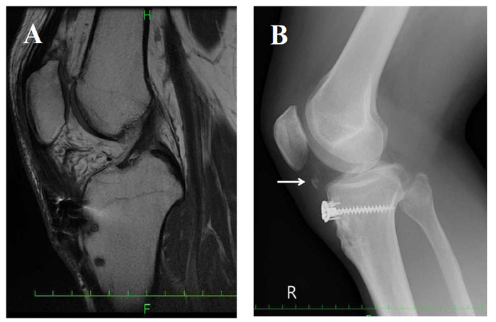
Fig. (4)
(A) Sagittal section of T2-weighted MRI and a lateral plain radiograph of the right knee at the six-month follow-up. (B) Twelve-month follow-up plain radiographs of the right knee. Bone union with ectopic patellar tendon ossification (small white arrow).