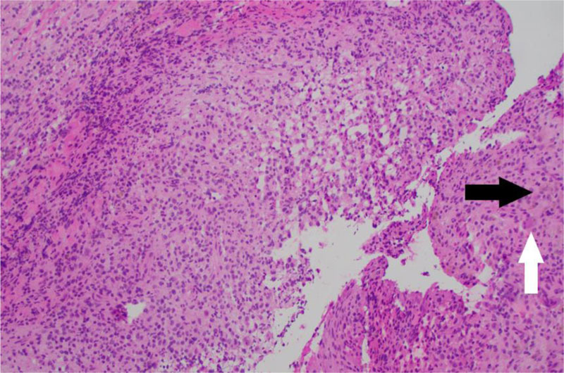
Fig. (3)
Frozen section pathology. Low power magnification shows moderately cellular tissue with admixed osteoclast-like giant cells (white arrow) and hemosiderin (black arrow) originally interpreted as ABC.