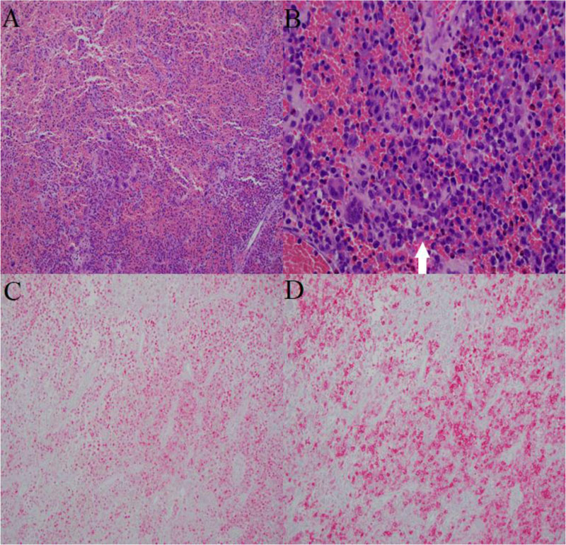
Fig. (4)
Final pathology. Low-power magnification shows loose histiocytic aggregates admixed with inflammation and hemorrhage (A). High-power image shows characteristic Langerhans cells (white arrow) with nuclear grooving and eosinophilic cytoplasm admixed with osteoclast-like multinucleated giant cells, and eosinophils (B). Langerhans cells show nuclear and cytoplasmic positivity for S100 (C) and characteristic membranous immunoreactivity for CD1a (D).