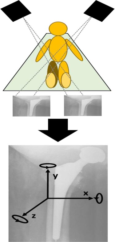
Fig. (1)
Radiostereometric analysis of the cementless femoral stem. Schematic drawing of radiographic examination of the operated hip with simultaneous exposure of two X-ray units. UmRSA calibration cage with X-ray plates under the examination table. Postoperative radiograph of a prosthesis with RSA tantalum markers on the implant and surrounding bone, coordinate system for RSA analysis.