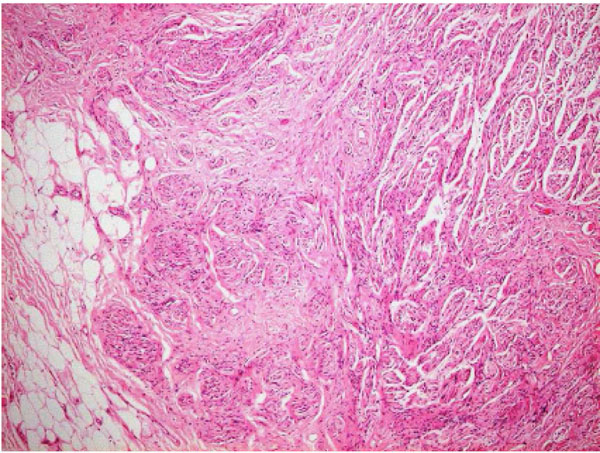
Fig. (3)
Histology of mass at x5 objective (H&E stain), showing nerves in disorganised fascicles, typical for the diagnosis of traumatic neuroma, plus some adjacent fat.