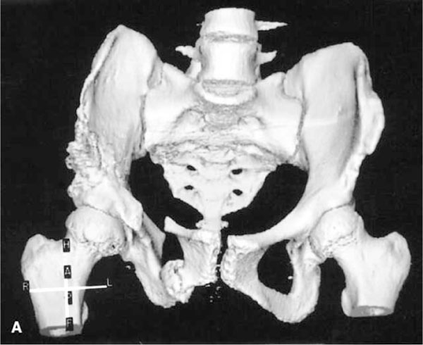
Fig. (2) A post-Steel osteotomy anterior view 3D-CT. The osteotomies of the superior pubic and ischial rami and from the ASIS to the sciatic notch are depicted. The bone graft has been removed from the superior wing of the ilium and repositioned to stabilize the acetabular fragment. Reproduced with permission from Frick S, Kim S, Wenger D; “Pre- and Postoperative Three-Dimensional Computed Tomography Analysis of Triple Innominate Osteotomy for Hip Dysplasia”, Journal of Pediatric Orthopaedics, Vol. 20, Issue 1, pp.116-23.