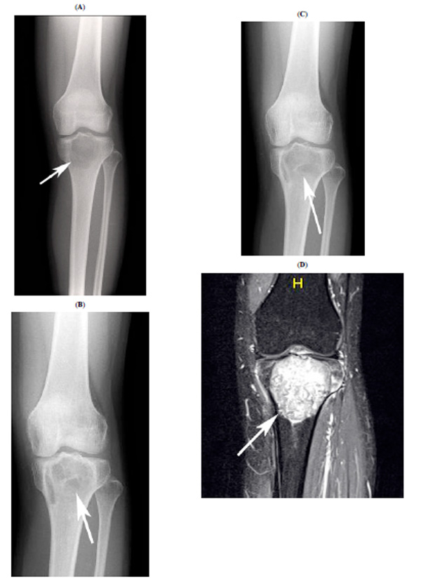
Fig. (2) Radiographic findings for case 1. This preoperative plain
radiograph shows the osteolytic lesion at the proximal tibia (A,
white arrow). Bone formation is apparent at 8 months after
curettage (B, white arrow). However, osteolytic change had
progressed at 16 months after the curettage. A plain radiograph
shows osteolytic lesions at 20 months after curettage (C, white
arrow). The osteolytic lesion was enhanced extremely by Gd in
MRI simultaneously (D, white arrow).