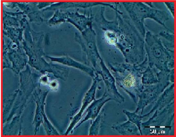
Fig. (2)
Growth of the chondrocytes was followed in flasks with invert microscope (under X40 magnification).