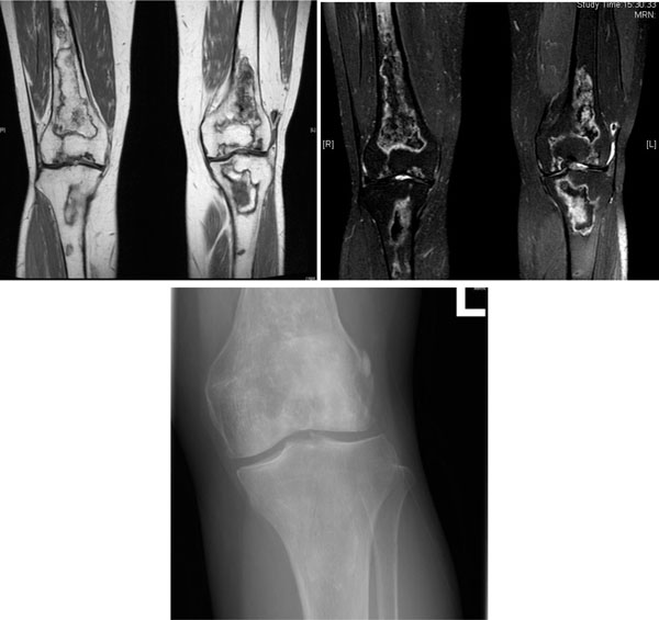
Fig. (2) AVN of both knees in a 53-year-old woman with SLE treated with glucocorticoid. (a) Coronal T1-weighted MRI scan. (b) Coronal
T2-weighted MRI scan. Multifocal serpiginous areas are present around both knees, from distal femoral shaft to proximal tibial shaft. The
areas have inner hypointense outer hyperintense rim on T2W sequences and enhancing outer rim. Similar lesion is also seen in left patella.
Overall features are suggestive of AVN which also involves the subarticular region. (c) X-ray left knee of the same woman. Increased
sclerosis is noted in subchondral region of lateral condyle, configuration of which is preserved. Serpigenous sclerosis is seen in medullary
region of proximal tibia and distal femur, suggesting bone infarct.