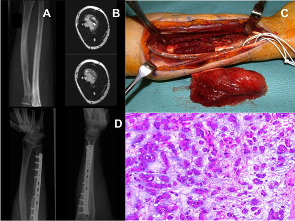
Fig. (1) (A-E) This radiograph was taken at initial presentation. The patient complained about a swelling which was tender to palpation, as well as paresthesia of the hand. It revealed a diffuse erosion in the ulna diaphysis with a broad zone of transition and adjacent cortical thickening (A). The MRI showed a soft tissue tumor with central signal enhancement (liquid or myxoid) and an intraosseous expansion (B). This shows the intraoperative situation with a segmental en-bloc resection of the ulna, before the allograft (fibula) and plate reconstruction was performed (C). Postoperative radiograph shows allograft incorporation and plate stabilization (D). The histological specimen revealed a low grademyxoidchondrosarcoma consisting of myxoid extracellular matrix with embedded cords and clusters of monomorph neoplastic cells (hematoxylin and eosin staining; original magnification 200x) (E).