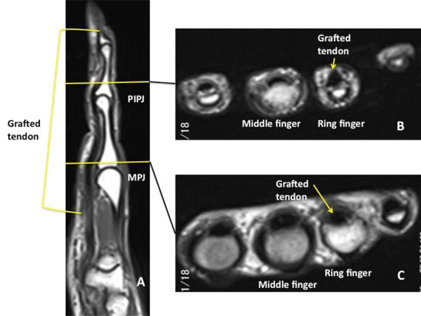
Fig. (4) Magnetic resonance imaging of the ring finger before tenolysis. (A) T1-weighted sagittal view showing the grafted flexor tendon
with low signal intensity. Cross-sectional view of the ring finger at the level of the proximal phalanx (B) and middle phalanx (C). The crosssectional
area of the grafted flexor tendon was similar to that of the adjacent digit.