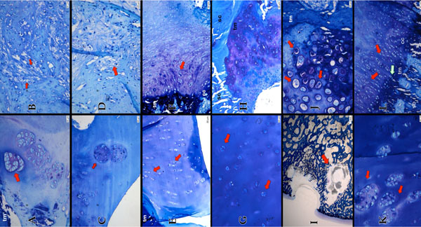
Fig. (3) Group 1 (VE) (Pictures A & B) huge cell nests (red arrow) in the adjacent cartilage and a slight loss of matrix staining visible. The
viability of the matrix is reduced. (tm=tidemark). (A) The defect part mainly consist of fibrous tissue (red arrows) (B) Group 2 (VECH)
(Pictures C & D): huge cell nests, but the viability of the matrix in the adjacent cartilage is maintained (C). Similar to Group 1 the Defect in
Group 2 is filled with fibrous tissue. Though it appears not as dense as in Group 1 (D) Group 3 (VECA) (Pictures E & F): The clusters in the
adjacent cartilage are visible in colums (red arrows) and there are only very few big cell nests (E). The defect part is mainly filled with a
mixture of fibro- cartilage (red arrow) and fibrous tissue (tm=tidemark, cc=calcified cartilage) (F) Group 4 (VECHCA) (Pictures G & H):
The adjacent cartilage appears almost normal hyaline like. Cluster appears as douplets (red arrow) huge cell nests are rare (not shown) (G)
Migration of cells from the subchondral bone into the defect part (sc=subchondral bone, tm=tidemark) (H) Group 5 (S) (Pictures I & J):
Cyste like lesion stretching into the adjacent cartilage (ground section 30-40 µm, staining with toluidine blue) (I) Hypercellular chondrocytes
(red arrows) at the defect basis (tm=tidemark) (J) Group 6 (CA) (Pictures K & L): Huge and numerous cell nests in the adjacent cartilage
(red arrows) (K) hyaline-like appearance in the defect part (red arrows). Chondrocytes grow from the subchondral bone into the defect part
(green arrow), similar as in Group 4 (cc= calcified cartilage, sb=subchondral bone, tm=tidemark) (L) (If not elsewise indicated: 5 µm
section, staining with toluidine blue).