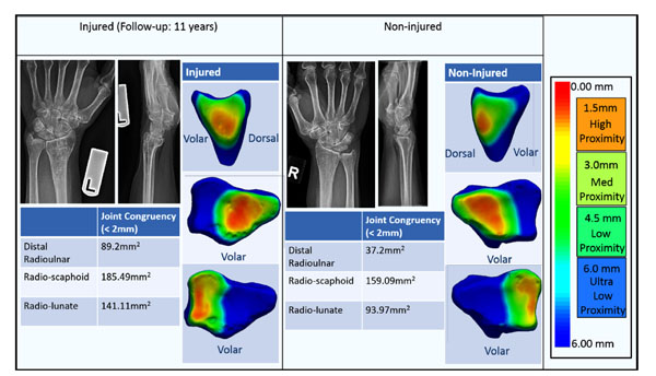
Fig. (3B) Case 2: Bilateral radiographs and joint congruency proximity maps are shown approximately 11years post fracture. A 60
year old female fell on her left wrist and suffered a full articular distal radius fracture that was treated with open reduction and internal
fixation. Radiographs show moderately distorted distal radius with a healed distal radius fracture that is slightly ulnar positive (+2.48mm) but
still within the acceptable ranges for ulnar variance (ASSH standards). Joint congruency maps appear to be asymmetric in the amount and
location of the regions of inferred high contact area between the injured and non-injured wrist. This region of high contact area is larger and
also located more centrally in the injured wrist (all three joints).