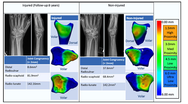
Fig. (3D) Case 4: Bilateral radiographs and joint congruency proximity maps are shown approximately 6 years post fracture. A 65
year old woman fell on her left wrist and suffered from a full articular fracture which was treated with external fixation and percutaneous
pins. Long term radiographs show a loss of volar tilt into dorsal angulation in the injured wrist. Her non-injured wrist also appears to be in
the neutral alignment in the sagittal plane but still within the acceptable alignment ASSH guidelines. Joint congruency maps appear to be
asymmetric in the amount and location of the regions of inferred high contact area between the injured and uninjured wrist. This region of
high contact area is larger and more distal-central in the non-injured distal radioulnar joint.