Fig. (5) (a) Axial T1-weighted spin echo and (b) axial T2-
weighted TSE fat suppressed MR images demonstrate the large
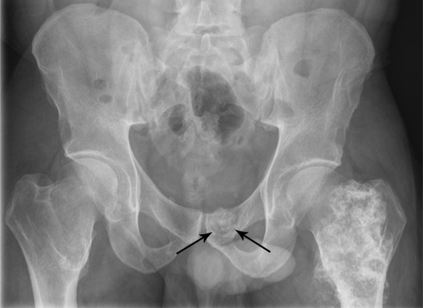
symptomatic osteochondroma arising from the posteromedial
aspect of the left proximal femur (short arrow) which displaces the
left gluteus maximus muscle. Also seen is a small osteochondroma
causing a pressure erosion on the posterior surface of the left pubic
body (long arrow). No bone marrow edema is associated with the
left pubic body erosion.