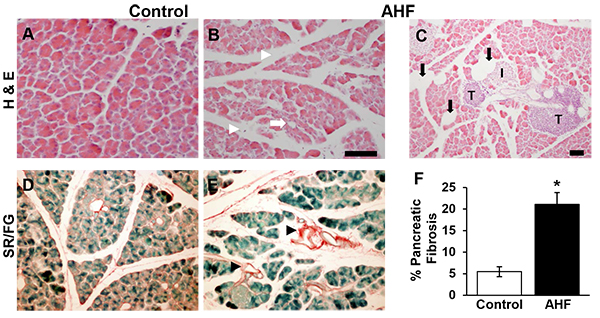
Fig. (1) Histopathology Indicative of Chronic Pancreatitis Is Evident in Tissue Sections from AHF Fed Mice. (A, D) Pancreatic tissue sections from control mice fed standard rodent chow show normal structure morphology. (B) Increased intralobular spaces (white arrowheads), degradation of acinar cells (white arrow), (C) adipocytes (black arrows), tumor-like structures, and (E) fibrosis (black arrowheads) in pancreas sections from AHF fed mice. (F) Quantification of fibrosis determined there was a significant increase in percent of the total tissue area stained with Sirius Red in pancreas of AHF fed mice compared to controls (p<0.05, two-tailed t-test). (I: islets of Langerhans; T: tumor like structures; * p<0.05; Scale bars, 100 μm).