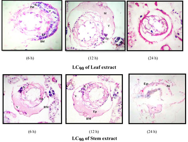
Fig. (5)
Photomicrographs of T.S. of midgut epithelium of early fourth instars of Aedes aegypti exposed to hexane extract of leaf and stem of Achyranthes aspera at LC90; Epithelial cells (Ep), Peritrophic Membrane (PM), Nucleus (N), Vacuole (V), Muscle (M) and Basement Membrane (BM) * Magnification: 40X.