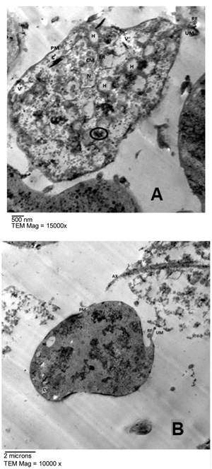
Fig. (3)
Transmission electron micrographs of untreated T. vaginalis were shown in with the plasma membrane (PM), the undulating membrane (UM) and its recurrent flagellum (RF) appear well preserved. In the cytoplasm, the nucleus (N), hydrogenosomes (H), vesicles (V), axostyle (AX) and costa (C) are observed. Few chromatin masses (CM) could be seen within the nucleus which is surrounded by endoplasmic reticulum (ER) and has a finely granular structure (Fig. A). Invagination of the plasma membrane forming coated vesicles (CV) is observed (Fig. B). Hydrogenosomes present a regular size from 0.3 to 0.5 mm, with a homogeneous matrix and could be seen in close contact with the axostyle. The hollow axostyle traverses the entire body of the parasite, protruding in the form of a spicule at its lower end. The walls of the axostyle consist of longitudinal filaments and its central cavity is filled with a granular mass (Fig. B). The costae have a specific structure resembling that of collagen fibers (Fig. A). Some glycogen granules (black circle) are observed (Fig. A).