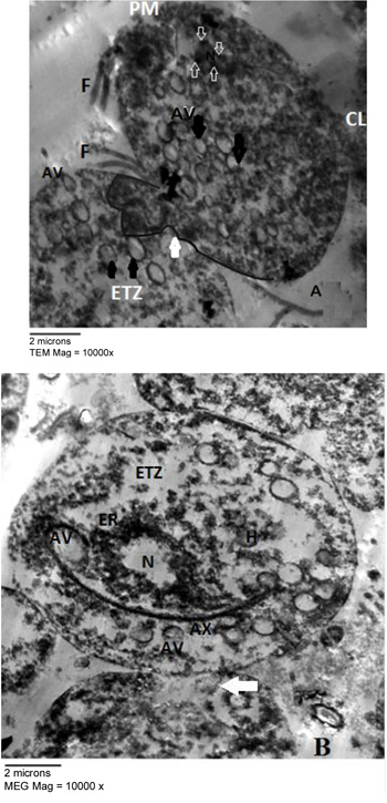
Fig. (4)
Transmission electron micrographs of Liagora farinose treated T. vaginalis showing adhesions between trophzoites which is mediated by numerous interdigitating pseudopodia (white compact arrow), damaged Plasma Membrane (PM), Cytoplasmic Leakage (CL), organelles disintegration with appearance of Electron Transleucent Zone (ETZ) (Fig. A), T. vaginalis trophozoites presented rounded Autophagic Vacuoles (AV) with different content (black arrows), some are located at the periphery of the cells and fusing with the plasma membrane, hydrogenosomes (H) are observed and chromatin accumulation inside nucleus (white vacuolated arrows) (Fig. A). Abnormal shape of nucleus (N) with widened Endoplasmic Reticulum (ER) is observed (Fig. B).