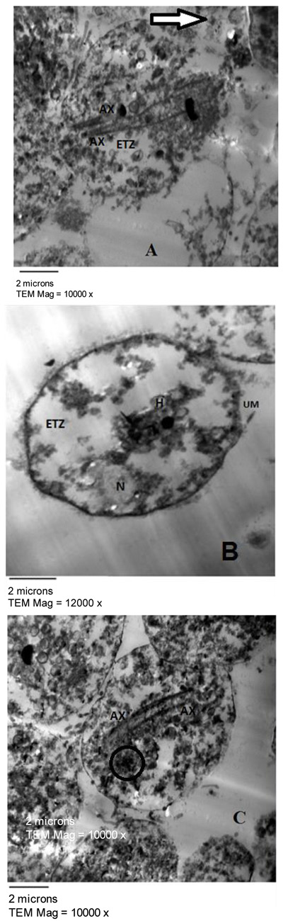
Fig. (6) T. vaginalis trophozoites treated with Holothuria fuscocinerea showing irregular ultrastructure and multiple axostyles (AX) (Figs. A and C) that appear dispersed in the cytoplasm. The content of the parasites was damaged seriously and the cell organelles mostly disappeared with the formation of extensive empty areas in the cytoplasm of the parasite, Electron Transleucent Zone (ETZ) (Figs. A and B) resulting in altered cell architecture. Large amount of glycogen granules (rosettes) (black circle) (Fig. C) are shown. Partially destructed cell membrane is observed. The vacuoles are destructed and hydrogenosomes are decreased in number (Fig. B) compared to untreated trophozoites. Cells showed washed out cytoplasm (white thick arrow in Fig. A).