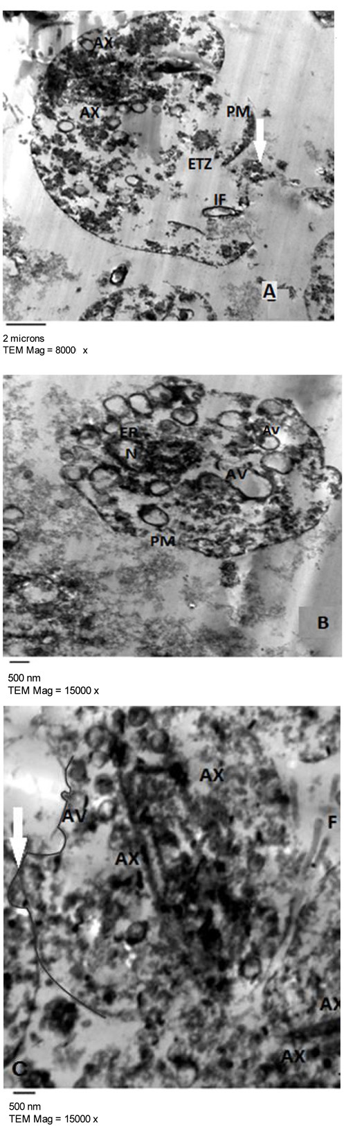
Fig. (8)
Transmission electron micrographs of metronidazole (1 µg/ml) showing T. vaginalis trophozoites with adhesions mediated by numerous interdigitating pseudopodia (white thick arrow, Fig. C), damaged Plasma Membrane (PM), Cytoplasmic Leakage (thick white arrow, Fig. A), organelles disintegration with appearance of ETZ (A). T. vaginalis trophozoites presented autophagic vacuoles (AV) with different content (Fig. B), some are located at the periphery of the cells and fusing with the plasma membrane (Fig. C). Nucleus is distorted in shape, chromatin is accumulated inside (Fig. B) and endoplasmic reticulum is widened (Fig. B). Internalized flagellae (IF) and multiple axostyles (AX) are seen (Fig. C).