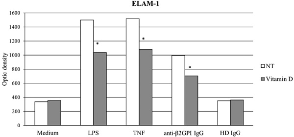
Fig. (1)
ELAM-1 expression in endothelial cells treated with APS IgG and vitamin D. Overnight pretreatment with vitamin D (1α,25-dihydroxy-vitamin D3, 1,25-OH-D3; SIGMA-Aldrich - Saint Louis, MI, USA) 80 nM significantly reduced the expression of Endothelial-Leukocyte Adhesion Molecule 1 (ELAM-1) induced by inflammatory stimuli (LPS and TNF-α) and anti-β2GPI IgG. No changes were detected in all other experimental conditions. Human umbilical vein endothelial cells (HUVECs) isolated from the umbilical cord vein were cultured in E199 (Gibco-Life Technologies - Groningen, The Netherlands), supplemented with 20% heat-injected Fetal Bovine Serum (FBS, PAA-GE Healthcare - Buckinghamshire, United Kingdom) in the presence of LPS (100 ng/mL, SIGMA-Aldrich - Saint Louis, MI, USA); TNF-α (40 ng/mL, R&D Systems – Minneapolis, MN, USA); or polyclonal IgG directed against β2GPI isolated from APS patient sera and IgG (200 μg/mL) from a pool of Healthy Donor (HD) sera respectively. IgG were isolated on a protein-G-Sefarose column (HiTrap Protein G, GE Healthcare Bio-Science AB, Uppsala, Sweden) as previously described by Tincani et al (97). A cyto-ELISA monoclonal IgG murine antibody was used to detect human ELAM-1 (R&D systems - Minneapolis, MN, USA). The choice of the doses and regimens was based on previous unpublished experimental data from our laboratory.anti-b2GPI: Anti-b2glycoprotein I; HD: Healthy Donor; LPS: lipopolysaccharide; NT: No Treatment; TNF: Tumor Necrosis Factor-alpha. (*p <0.05)