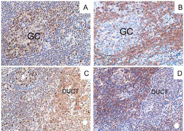
Fig. (2). BAFF and BAFF-R expression in parotid gland (MESA pre-rituximab in the Panel A and C, and MALT NHL postrituximab in the Panel B and D) during the follow-up Microphotograph showing diffuse BAFF (A and B) and BAFF-R (C and D) expression in the salivary gland of the patient in the parotid biopsies relative to MESA 2002 (A and C) and post-rituximab MALT NHL 2005 (B and D). BAFF strongly positive cells (brown) were detected in the MESA both inside the infiltrated areas (germinal centres=GC in panel A) and in the ducts (data not shown), but intense BAFF expression with the same pattern were detected also in the B-cell MALT NHL despite the treatment with rituximab (panel B). Sequential sections show strong expression of BAFF-R in mantle zone B cells (panel C for the MESA), and in the infiltrating lymphocytes surrounding the epithelial structures (panel D for the MALT NHL) (Original magnification 100x).