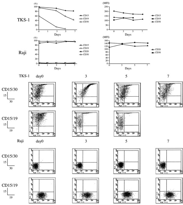
Fig. (2) Effects of PMA on surface makers of TKS-1 and Raji cells. TKS-1 and Raji cells (5 x 10⁵ cells/ml each) were resuspended in fresh medium containing either 10 ng/ml PMA or solvent. Two-thirds of the medium was replaced with fresh medium every 2 days. Expression of CD15, CD30, CD19, and CD20 was examined by flow cytometry at 0, 1, 3, 5, 7 days after starting the experiment. Data are representative of three repeated experiments. Dot plots show representative flow cytometry data.