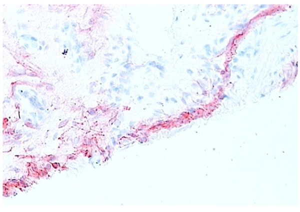
Fig. (5)
Normal synovium (X200 magnification) stained for CD55
+
fibroblasts (red). Synovial fibroblasts make up most of the cells in the lining layer in the normal synovium intimal layer in contrast to RA synovial tissue (contrast with Figs.
4
,
7
).