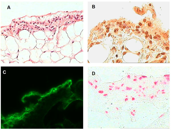
Fig. (1) Synovial tissue analysis of biopsy material obtained from a symptomatic knee joint shortly after onset of articular symptoms. A) H&E light microscopy showing marked disruption of the synovial lining cell layer. The lining cells appear to be floating in an amorphous extra-cellular matrix. The sublining stroma is relatively unremarkable with minimal evidence of inflammatory infiltration. B) Intracellular citrullinated antigens are detected by intense staining of the synovial lining cells using a polyclonal anti-citrulline antibody. There is less intense staining of the surrounding matrix. C) Immunofluoresence staining of synovial tissue for C3 showing positive staining in the lining layer. Similar results were seen with IgG, IgA, and fibrin staining. D) The synovial lining cells exhibit evidence for widespread apoptosis as detected by TUNEL staining. Original magnification x 200 for A), and x 400 for B), C), D).