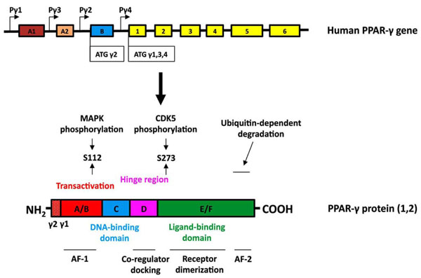
Fig. (1) Structure of the human PPARγ gene and protein. (Upper panel, gene structure) Expression of PPAR-γ involves differential
promoter usage, and the relative positions of the four known PPAR-γ promoters are designated as Pγ1-Pγ4. The transcript variants γ1, γ3,
and γ4 encode the PPAR-γ1 isoform. Exon B (blue box) encodes the 28 additional amino acids found at the amino terminus of human PPAR-γ2 (30 amino acids in mouse PPAR-γ2). Exons 1–6 (yellow boxes) are common in all PPAR-γ1 transcripts and when they are spliced to exon
B encode full-length PPAR-γ2. The sizes of the exon boxes approximate the relative lengths of each exon; however, the introns (depicted as
straight lines) are not drawn to scale. (Lower panel, protein structure) The hypervariable A/B domain (red box) contains the activation
function-1 (AF-1) domain. Human PPAR-γ2 contains a 28 amino acid amino terminal region. The C-domain (blue) contains the DNA
binding domain (DBD). The D domain (hinge region, pink box) allows for conformational change following ligand binding to promote
coregulator (coactivator or corepressor) docking. The E/F region (green box) contains the ligand binding domain (LBD) of PPAR-γ and the
activation function-2 (AF-2) domain. PPAR-γ can be phosphorylated by MAP kinases at S112 or by CDK5 at S273. The AF2 domain
participates in ubiquitin-dependent degradation and is necessary for full ligand-induced PPAR-γ transcriptional activity.