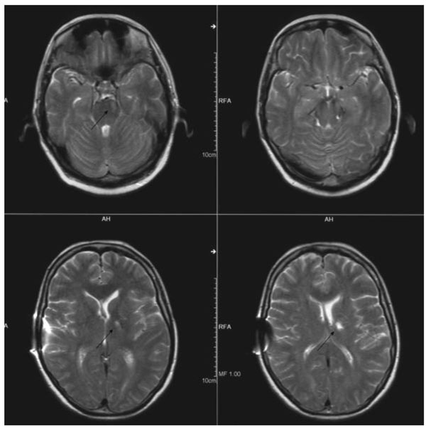
Fig. (4)
Cerebral MRI six months after bleeding: ischemic lesions in left thalamus, left pons and mesencephalon (arrows).