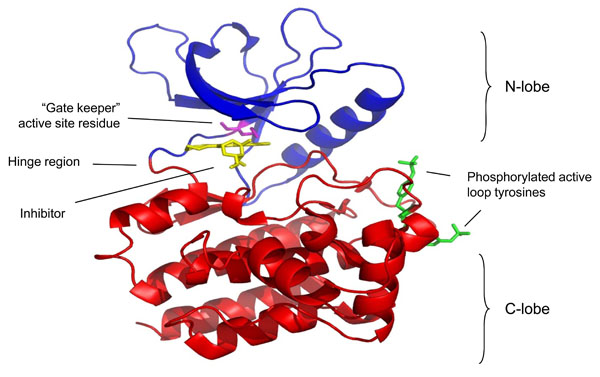
Fig. (2) Crystal structure of the Jak3 kinase domain in complex with staurosporine (pdb accession code 1YVJ). This structure
captures the active conformation of Jak3 with both active loop tyrosine residues phosphorylated (green). The molecule can be described in
two halves, with the N terminal lobe presented in blue and the C terminal domain in red. These are linked by a hinge region that forms part of
the active site. Highlighted in magenta within the active site is the gate keeper residue. Bound within this site is an analogue of the inhibitor
staurosporine, and its proximity to the “gate keeper” residue highlights why this residue and this region are critical for the specificity of
inhibitors for individual protein kinases.