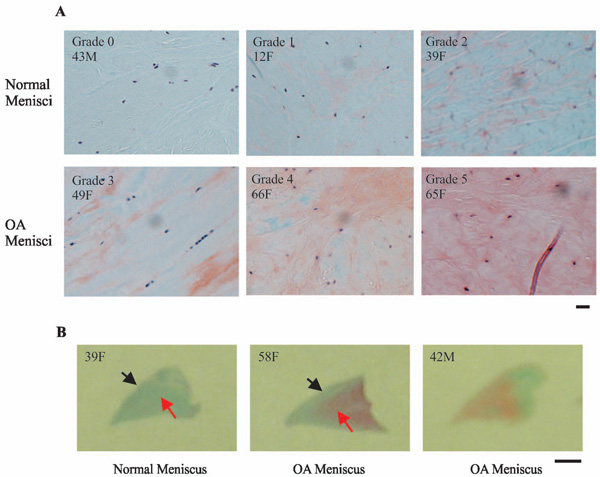
Fig. (6) Representative images of safranin-O staining. (A) Normal menisci (horizontal sections of the central portion of menisci) displayed
minimal or weak orange-red staining (top photos). In contrast, OA menisci (horizontal sections of the central portion of menisci) displayed
moderate to very strong orange-red staining (bottom photos). Scale bar: 200 µm. (B) Normal menisci (transverse sections of the entire
meniscus) displayed minimal orange-red staining. OA menisci (transverse sections of the entire meniscus) displayed moderate to strong
orange-red staining. The increase of proteoglycan staining in OA menisci compared to normal menisci was more prominent in the middle
and deep zones than that in the surface zone. Scale bar: 2.5 mm.