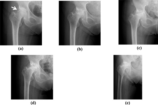
Fig. (1) (a)
At the first visit, a plain radiograph of the right hip showed joint space
narrowing (KL grading II). A laddering-shaped deformity in the right lateral
proximal femoral head and fracture-like line were observed (white arrow). (b)
Eight months after onset, a plain radiograph revealed increased joint space
narrowing, a band around the necrotic area, and a concave shape to the right
femoral head. (c) The necrotic region of the right femoral head became
noticeably worse a year after onset. (d) The right femoral head had
collapsed at 3 years after onset. (e) A recent radiograph showed
progressive destruction of the right femoral head and the remaining joint line
exhibited osteosclerotic change. Note that the patient did not report any joint
pain.