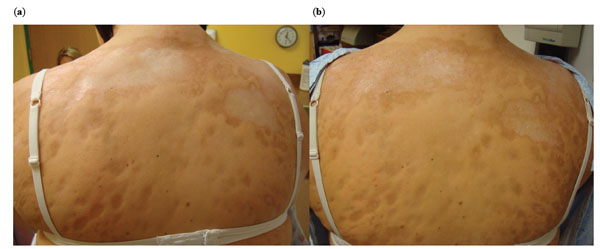
Fig. (2) Pre- and Post-treatment photographs of the upper back. The initial back lesions (a, left panel) demonstrate yellow/white waxy
lesions with thick skin coalescing over her upper back. Post-treatment photographs (b, right panel) demonstrate decreased white/yellow waxy
appearance of lesions, with less uniform skin thickness and ‘breaking up’ of lesions.