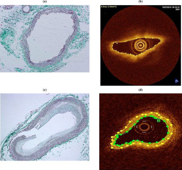
Fig. (3) Cross-section of a medium-sized pulmonary artery from a patient without pulmonary hypertension showing a medium-sized
pulmonary artery without hypertensive changes (Masson Trichrome, x 100) (a). OCT image of the same cross-section where no fibrosis is
appreciated (b). Cross-section of a medium-sized pulmonary artery from a patient with pulmonary hypertension showing severe intimal
fibrosis (Masson Tricrome, x 100) (c). OCT image of the same cross-section where the area of fibrosis has been marked ( area between green
and yellow lines ) (d).