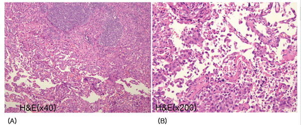
Fig. (4) Histological findings of cervical lymph nodes. (A) Lymph nodes are almost replaced by angiosarcoma, which forms an
anastomosing network of sinusoids, lined by hyperchromatic, nucleated endothelial cells (hematoxylin and eosin (H&E) staining, × 40). (B)
A distinct sinusoid pattern with malignant endothelial cells, some of which are scattered in the lumen of sinusoids, is seen (H&E stain, x
200).