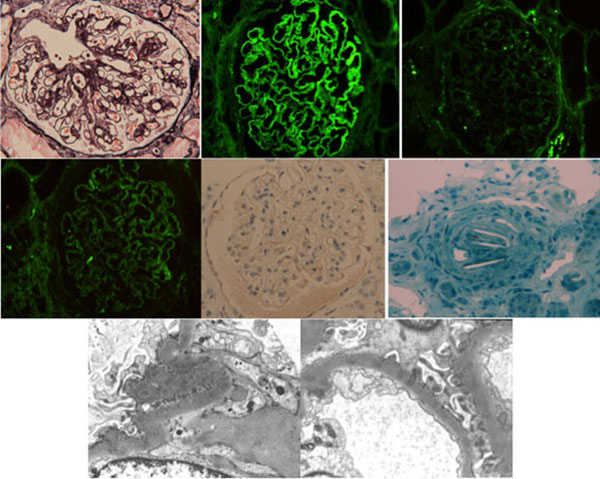
Fig. (1)
(Top) Light photograph and confocal microscopy images of glomeruli showing diffuse thickening of glomerular basement membranes with some spikes (periodic acid–methenamine silver stain) and immunofluorescence staining for immunoglobulin G (IgG), C3, and C1q (×400). IgG4 staining was negative in paraffin section (×400). Methylene blue staining of a 1-µm-thick Epon-embedded section showing one occluded interlobular artery containing a pathognomonic, biconvex, needle-shaped cleft, which indicates a cholesterol crystal (×400). (Bottom) Electron micrographs showing focal effacement of podocyte foot processes, fine electron-dense deposits in the mesangial matrix, and small subepithelial electron-dense deposits in the loops (A, ×6000; B, ×8000).