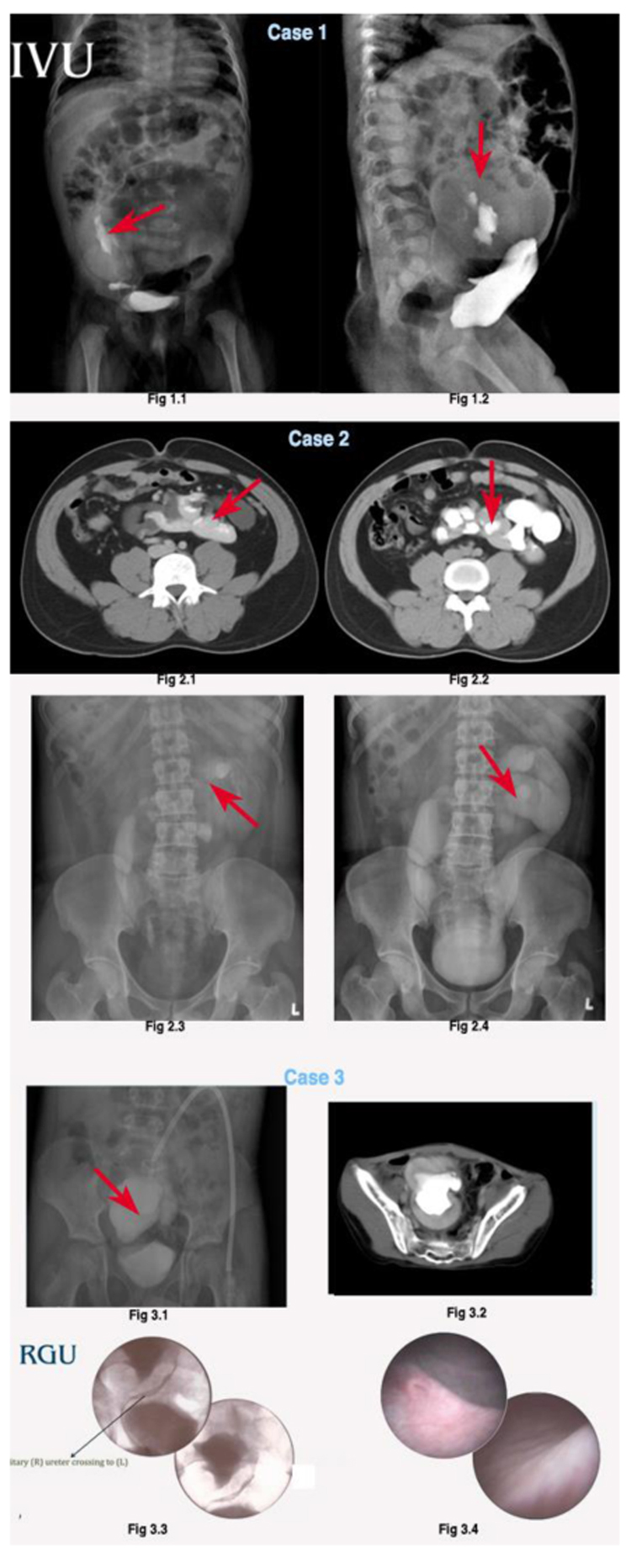
Fig. (1) 1.1,1.2: IVU showing right to left superior ecopia, right hydroureteronephrosis with left hydronephrotic unit with atretic ureter. 2.1, 2.2, 2.3,2.4: IVU/CECT showing bilateral moderate hydroureteronephrosis with right to left ‘L’ shaped fusion 3.1,3.2: IVU/CECT KUB revealed a right to left cake shaped fusion. 3.3: RGP showed right solitary ureter crossing to the left side 3.4: On cystoscopy, left hypoplastic ureteric orifice was present.