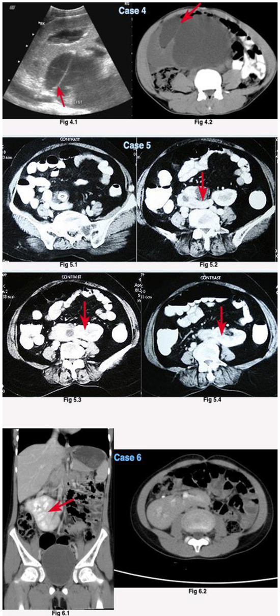
Fig. (2) 4.1, 4.2: USG and CECT showed right to left disc shaped fusion, (R) hydronephrotic kidney fused with (L) grossly hydronephrotic unit. 5.1,5.2,5.3,5.4: CECT KUB showing a left to right crossed L shaped fusion ectopia with a right renal pelvic calculi measuring 2cm with (R) unit hydronephrosis. 6.1, 6.2: showed a left to right sigmoid ‘S’ shaped crossed fusion ectopia.