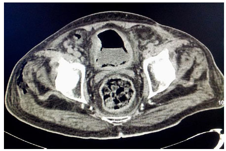
Fig. (2)
Axial noncontrast CT scan study at the level of urinary bladder demonstrates partially distended urinary bladder with thickened walls and perivesical fat stranding with intraluminal and intramural pockets of air, consistent with emphysematous cystitis. Mottled pockets of air involving right tensor fascia lata and adjacent gluteus maximus muscles. Reactive inflammatory changes or collection are minimal due to poor immune response.