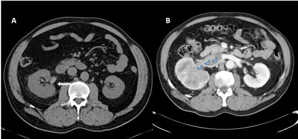
Fig. (1) (A) Non-contrast CT scan of abdomen and pelvis demonstrates slightly enlarged right kidney with mild perinephric fat stranding.
Note hyperdense blood in the pelvicalyceal system consistent with hematuria (marked by white arrow). (B) Contrast enhanced CT scan of the
abdomen and pelvis reveals an infiltrating mass involving the whole kidney with dilated and thrombosed right renal vein (marked by
chevrons).