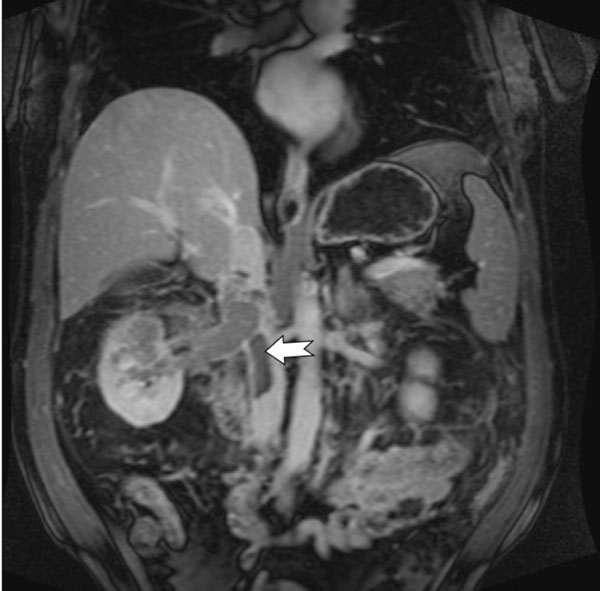
Fig. (2)
Contrast enhanced fat saturated T1 weighted coronal MRI image demonstrates infiltrative right renal mass with extension of the tumor thrombus into the right renal vein and the inferior vena cava (marked by notched arrow).