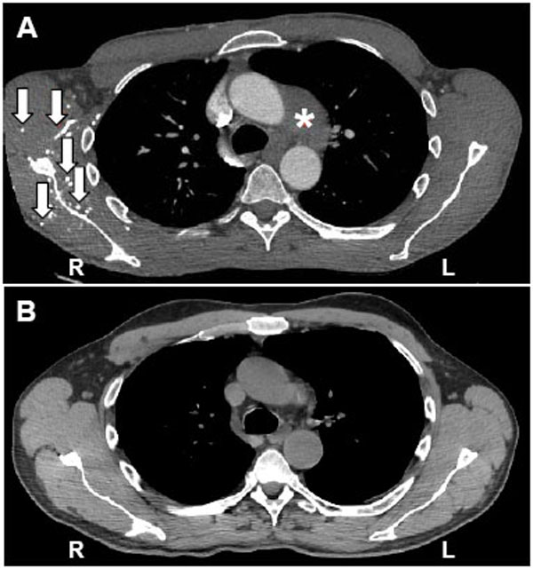
Fig. (1) Computed tomography scans of the chest. A nonenhancing soft-tissue mass in the aortopulmonary window (*) is seen on a CT angiogram of the chest (A). Extensive venous collateral circulation in the chest wall (arrows) indicates central venous stenosis or occlusion (A). A decrease in the size of softtissue mass in the aortopulmonary window four months after the initiation of immunosuppressive therapy on a non-enhancing highresolution CT scan of the chest (B). There is no evidence of fibrosis in the mediastinal fat (B).