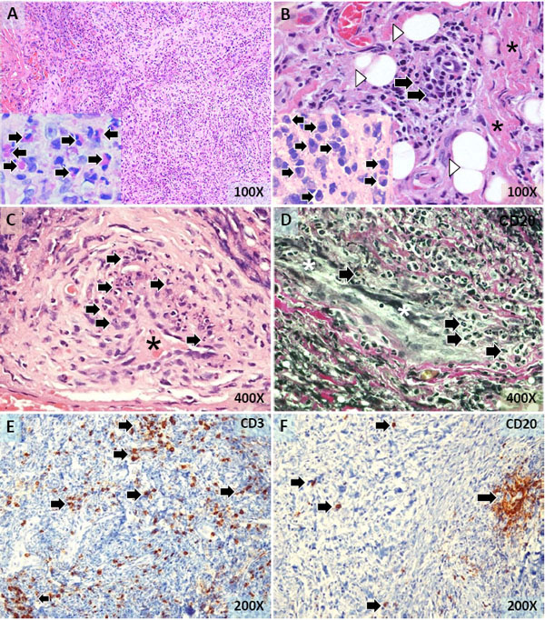
Fig. (2) The histology of the mediastinal mass. Fibrous connective tissue containing numerous mononuclear and polymorphonuclear inflammatory cells (A). Numerous eosinophils (arrows in the insert) were present (A). A focus of inflammatory cell infiltration (arrows),fibrous connective tissue (*), and scattered residual fat cells (arrowheads) (B). Numerous plasma cells (arrows in the insert) were observed (B). Small blood vessels (*) with mononuclear and polymorphonuclear inflammatory cells (arrows) in the wall indicative of small-vessel vasculitis (C,D). Numerous T lymphocytes (arrows) scattered throughout the lesion (E). B lymphocytes forming a small irregular aggregate (arrowhead) and individually dispersed (F). Tissue sections were stained with hematoxylin-eosin (A-C) and elastic Van Gieson (D). Tissue sections were also immunostained for CD3 (E) and CD20 (F). Magnifications are as indicated.