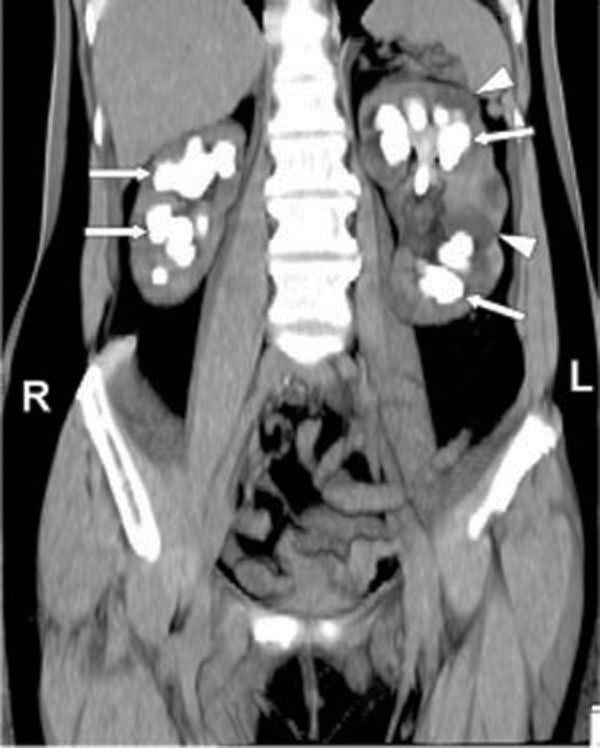
Fig. (1) Bilateral staghorn calculi in a 50 year old female patient
with recurrent urinary tract infection. Coronal maximum intensity
projection non contrast CT scan image shows calculi filling the
renal calices (arrows). Dilated calices are also present in the left
kidney (arrowheads).