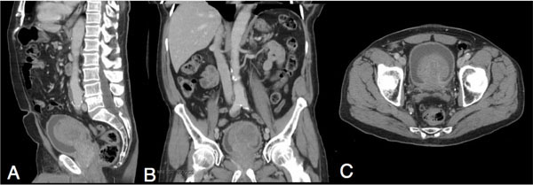
Fig. (1)
Representative cuts from a computed tomography (CT) scan of patient’s abdomen and pelvis showing an 8-centimeter mass from the prostate gland extending into the lumen of the bladder. (A) Sagittal view (B) Coronal view (C) Transverse view.