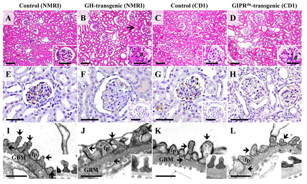
Fig. (13)
Glomerular morphology (A-D), detection of glomerular
podocalyxin (PODXL) abundance by immunohistochemistry (IHC, E-H), and
electron-microscopic demonstration of anionic sites on the surface of
glomerular podocytes by polyethyleneimine (PEI) labeling (I-L) in a GH-transgenic
mouse (male, six week-old, NMRI background, B, F, J), a GIPRdn-transgenic
mouse (six-month-old, CD1 background, D, H, L), and associated
age-matched control mice of the respective genetic background (A, E, I,
and C, G, K). A-D: The GH-transgenic and the GIPRdn-transgenic mouse
display glomerular hypertrophy and mesangial expansion and matrix accumulation
(#). Additional lesions in the GH-transgenic mouse comprise glomerular
capillary hyalinosis, synechiae between the glomerular capillaries and the
parietal epithelium of the Bowman capsule (*), and tubular protein casts (open
arrow in B). E-H: Compared to control mice (E, G), the immunohistochemical
PODXL staining intensity (brown color) at the surface of glomerular podocytes is
reduced in some glomeruli of the GH-transgenic mouse (F) and the GIPRdn-transgenic
mouse (H). Insets to F and G: IHC negative controls. A-H: Paraffin sections, H
& E staining (A-D). For IHC (E-H), DAB was used as chromogene (brown color),
and hemalaun as nuclear counterstain. Bars = 50 μm, and =10 μm in insets. E,
F: Transmission electron-microscopic (TEM) detection of anionic sites on
podocyte surfaces with the cationic probe PEI. In non- transgenic control mice
(I, K), the podocyte foot processes (fp) and the outer and inner laminae rarae
of the glomerular basement membrane (GBM) display regular, punctate,
electron-dense labeling patterns (arrows). In comparison, the broadened
podocyte foot processes in some glomeruli of the GH-transgenic and the GIPRdn-transgenic mouse show a slightly decreased number of PEI labeled anionic sites.
Insets: Podocyte foot processes of unlabeled control samples. Bars = 0.5 μm.