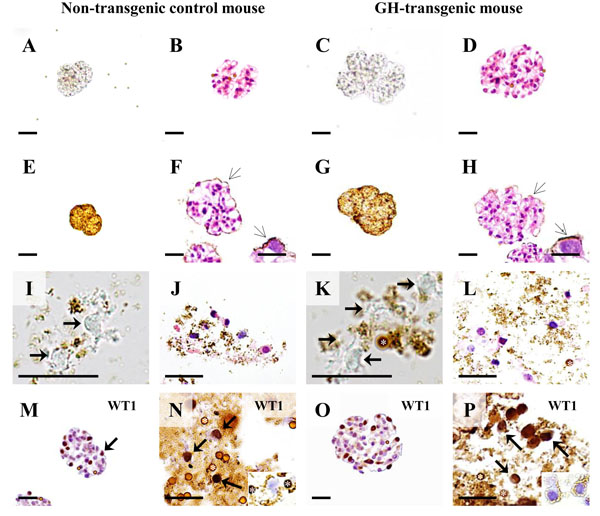
Fig. (14)
Podocyte isolation in a growth hormone (GH) transgenic
mouse and non-transgenic control mouse (male, six-weeks of age). A-D:
Isolated glomeruli. A, C: Native, unstained preparations. B,
D: H & E stained paraffin sections. E-H: Isolated
glomeruli after coating with cationic colloidal silica-coated ferromagnetic
nanoparticles (NPs, open arrows). E, G: Native, unstained
preparations. F, H: H & E stained paraffin sections. Arrows mark
NPs on the glomerular surface. Insets: Detail enlargement of NP-covered (open
arrows) podocytes. Note the larger sizes and section profiles of the glomeruli
of the GH-transgenic mouse, as compared to the control mouse. I-L:
Podocyte isolates. Asterisks mark DynabeadsTM. I, K: Native,
unstained preparations. Arrows mark podocytes. J, L: H & E
stained paraffin sections. M-P: Immunohistochemical detection of
the podocyte marker protein Wilms tumor 1 (WT1) in paraffin sections of isolated
glomeruli (M, O) and paraffin sections of podocyte isolates (N, P). Podocytes
(arrows) display nuclear WT1-immunoreactivity. Insets to N and P: Podocyte
profiles in IHC negative control sections. Asterisks mark DynabeadsTM. Bars in
A-H = 50 μm, and = 10 μm in insets. Bars in I-P = 25 μm.