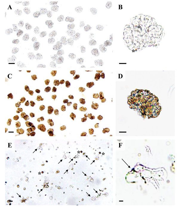
Fig. (6)
Light microscopy of podocyte isolation procedure steps.
A, B: Isolated glomeruli with light-brown magnetic DynabeadsTM inside
glomerular capillaries (asterisks in B). C, D: Isolated glomeruli
after incubation with a 10 mg/ml suspension of cationic colloidal silica-coated
ferromagnetic nanoparticles (NPs) in coating buffer and treatment with PAA. The
NPs form a dense coating of the glomerular surface. E: Podocyte isolate
with podocytes (arrows) and free magnetic DynabeadsTM(asterisks). F:
Detail enlargement of an isolated podocyte with NP’s on the cell surface
(arrow). Note the primary processes of the podocyte. Native, unstained
preparations. Bars = 50 μm (A, C), 20 μm (B, D), 10 μm (E), 5 μm (F).