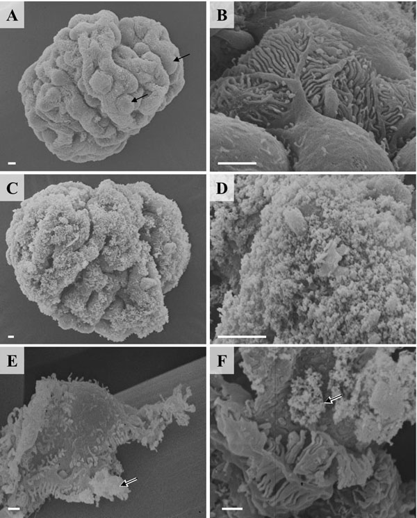
Fig. (8)
Scanning electron microscopy (SEM) of podocyte isolation
procedure steps. A, B: Isolated glomerulus. Podocytes are marked by arrows
B: Podocyte on a peripheral glomerular capillary. Note the interdigitating
foot processes of neighboured podocytes. C: Isolated glomerulus after
coating with cationic colloidal silica-coated ferromagnetic nanoparticles
(NPs). D: Detail enlargement of C. E: Isolated podocyte with NP’s
on the cell surface (arrow). Note that some NPs will have detached from the cell
surface during processing of the sample for SEM. F: Detail enlargement
of foot processes of isolated podocytes. Arrow marks NPs. Bars = 1 μm.