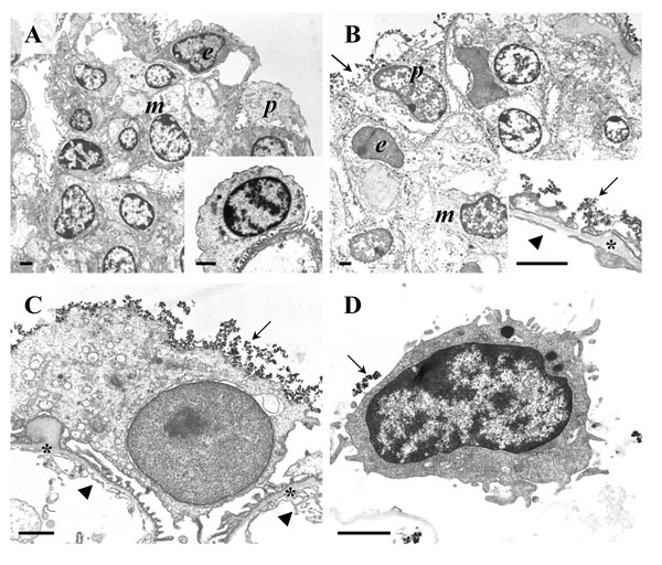
Fig. (9)
Transmission electron microscopy (TEM) of podocyte
isolation procedure steps. A, B: Isolated glomeruli. Peripheral podocytes (p),
endothelial cells (e), and mesangial cells (m) are indicated. A:
Isolated glomerulus before coating with cationic colloidal silica-coated
ferromagnetic nanoparticles (NPs). The inset to A shows a detail enlargement of
a podocyte on a peripheral glomerular capillary. B: Isolated glomerulus
after coating with NPs. The inset to B shows a detail enlargement of a
peripheral glomerular capillary wall. C: Detail enlargement of a
peripheral podocyte in a glomerulus coated with NPs. In B and C, the
glomerular basement membrane (GBM) is indicated by asterisks. NPs attached to
the podocyte surface, respectively the surface of podocyte foot processes are
marked by arrows. Arrowheads indicate the fenestrated endothelium. Note the
absence of NPs from the GBM and from endothelial and mesangial cell surfaces.
D: Isolated podocyte with NP’s on the cell surface (arrow). Note that some
NPs will have detached from the cell surface during processing of the sample
for TEM. Bars = 1 μm.