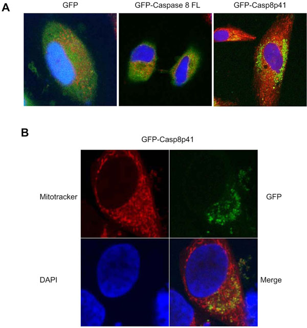
Fig. (4) Casp8p41 colocalizes with mitochondria. (A) HeLa cells were transfected with GFP, GFP-casp8p41, or GFP-casp8FL. After 6 hours of transfection, cells were stained with MitoTracker® red, and nuclei was counterstained with DAPI. Staining was visualized by confocal microscopy. The figure shows the overlay of the staining: GPF constructs (green), Mitotracker (red), and DAPI (blue). (B) Same as in A. Images of GFP-casp8p41 transfected HeLa cells are shown individually for each staining and as an overlay.